45 lower extremities labeled
Leg Anatomy - FPnotebook.com The limb that is composed of the hip, thigh, leg and foot. Definition (MSH) The region of the lower limb in animals, extending from the gluteal region to the FOOT, and including the BUTTOCKS; HIP; and LEG. Concepts. Body Location or Region ( T029 ) MSH. D035002. SnomedCT. 61685007, 116013008. Anatomy Of The Lower Extremity Veins - Varicose Veins Valves in superficial veins of the lower extremity usually are located near to the termination of major tributaries. Some valves are well developed with marked sinusoid dilation at their base, others are more delicate in their structure. In the GSV there are about six valves, with more valves located below than above the knee.
The Lower Extremity: The Knee, Ankle, and Foot | Kinesiology ... Analyze the fundamental movements of the lower leg and foot with respect to joint and muscle actions. Describe the common injuries of the leg, knee, and ankle. Perform an anatomical analysis of the lower extremity in a motor skill.

Lower extremities labeled
Anatomy, Shoulder and Upper Limb, Arm Structure and Function It consists of three sections, the upper arm, forearm, and hand. It extends from the shoulder joint to the fingers and contains 30 bones. It also consists of many nerves, blood vessels (arteries and veins), and muscles. The nerves of the arm are supplied by one of the two major nerve plexus of the human body, the brachial plexus. Bones of lower limbs Bone: Lower limb -. Bones of lower limbs. ○Including bones of pelvic girdle (hip bones) and lower limb. ○compared with upper limb.70 pages Lower extremity: MRI anatomical atlas - e-Anatomy - IMAIOS This cross-sectional human anatomy atlas of the lower limb is an interactive tool based on MRI axial images of the human leg. Anatomical structures of the lower limb (hip, thigh, knee, leg, ankle and foot) and specific regions (compartment of the lower limb) are visible on dynamic labeled images. Cross sectional anatomy of the hip : axial slice ...
Lower extremities labeled. Muscles of the lower limb | Radiology Reference Article - Radiopaedia The muscles of the lower limb are numerous and complex. Their origins and insertions are difficult to remember, and they are best considered as parts of general functional groups. iliopsoas psoas major psoas minor iliacus buttocks gluteal region gluteal muscles gluteus maximus gluteus medius gluteus minimus tensor fasciae lata lateral rotator group Upper limb anatomy: Bones, muscles and nerves | Kenhub The shoulder joint is reinforced with two groups of muscles, superficial and deep. Superficial muscles include the deltoid and the trapezius, whereas the deep group contains the supraspinatus, infraspinatus, teres minor and subscapularis (rotator cuff) muscles. To easily remember the rotator cuff muscles, use the mnemonic given below! Anatomy, Bony Pelvis and Lower Limb, Femoral Artery The femoral artery is a large vessel that provides oxygenated blood to lower extremity structures and in part to the anterior abdominal wall. The common femoral artery arises as a continuation of the external iliac artery after it passes under the inguinal ligament. The femoral artery, vein, and nerve all exist in the anterior region of the thigh known as the femoral triangle, just inferior to ... The Biomechanics of the Human Lower Extremity | Basic Biomechanics, 7e ... In contrast, the lower extremity is well equipped for its functions of weight bearing and locomotion. Beyond these basic functions, activities such as kicking a field goal in football, performing a long jump or a high jump, and maintaining balance en pointe in ballet reveal some of the more specialized capabilities of the lower extremity. This ...
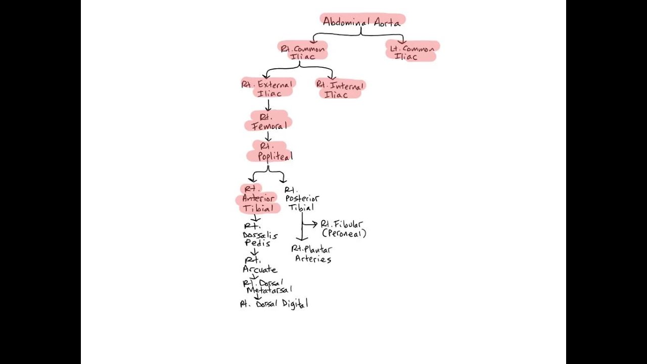
Treatment of Varicose Veins of the Lower Extremities The Clinical-Etiology-Anatomy-Pathophysiology (CEAP) classification serves as a basis to categorize the clinical presentation of the patient, ... The analysis of evidence reviewed for the treatment of varicose veins of the lower extremities included a multicenter randomized controlled trial, prospective study, observational study, retrospective ... Labeled imaging anatomy cases | Radiology Reference Article ... This article lists a series of labeled imaging anatomy cases by body region and modality. Brain CT head: non-contrast axial CT head: non-contrast coronal CT head: non-contrast sagittal CT head: angiogram axial CT head: angiogram coronal CT... Anatomy of the lower extremity arteries and bones ... - IMAIOS Anatomy of the arteries and bones of the lower limb based on 3D pictures and angiogram (angiography). This part of the interactive atlas of anatomy of the human body is about the arterial vasculature of the pelvic girdle, pelvis, thigh, knee, leg and foot and the study of bones and joints. It includes a 3D reconstruction of bones and arteries ... Muscles Of The Lower Extremity 1 - ProProfs Quiz Welcome to the electronic human anatomy and physiology classroom of the 21st Century. This test will focus on the muscles and muscle groups of the lower extremity of the thigh, lower leg, and foot. The musculature, bony structure, and joints of the pelvic girdle function in locomotion and maintenance of stability. Please note the terms "action" and "function" have the same meaning. You will ...
Lower Extremity Landmarks - TeachMe Orthopedics Lower Extremity Landmarks By admin On Jan 4, 2022 DERMATOMES MYOTOMES OSTEOTOMES SCIATIC NERVE Inferior division of lumbar L4, L5 and sacral S1, S2, S3 nerves. Emerges from the greater sciatic foramen. Lies below the piriformis muscle (m.), deep to gluteus maximus m. on the posterior wall of the pelvis. Vascular Anatomy of the Lower Extremity: A Practical Guide to Vascular ... Fig. 8.7 The lower extremity can be divided into four anatomic subregions: (1) gluteal, (2) hip and thigh, (3) knee and leg, and (4) ankle and foot. Within these regions, there are 21 cutaneous vascular territories. See Table 8.1 to explain acronyms. 8.8 Regional Anatomy of Perforators of the Lower Extremity Radiological anatomy of the lower limb - e-Anatomy - IMAIOS Anatomy of the lower extremity in standard radiology. This radioanatomy module of the lower limb presents 24 conventional radiographs with 192 anatomical structures labeled. It is particularly useful for radiologists, electroradiology students, emergency physicians, orthopedic surgeons and rheumatologists, but may be used on a daily practice or ... Arterial vascular anatomy on the lower limb (DSA) - IMAIOS Some terms were added, commonly used for the radiological vascular anatomy of the lower extremities, such as "superficial and common femoral artery" or "tibiofibular trunk". We would like to thank Michel Alauzen M.D. (Vascular surgery, Montpellier - France) for his precious advice in vascular anatomy. Bibliography:
Skeletal System Diagrams
Lower Extremity Anatomy: Parts and Functions | New Health Advisor The famous 'Achilles' tendon is the most noteworthy structure in your lower extremity anatomy. It joins three muscles: calf, soleus and plantaris to your heel bone. The Achilles tendon stores all the essential elastic energy required for jumping, running and several other physical activities. 4. Nerves
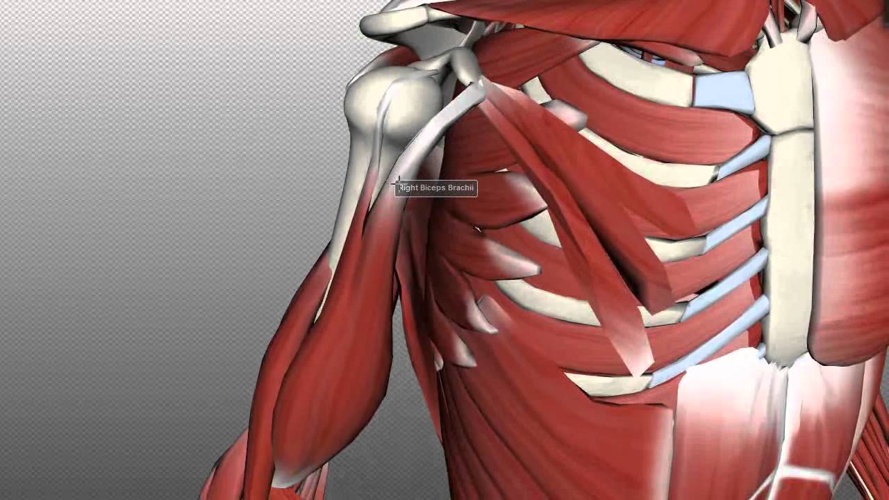
Muscles of the Upper Arm - Anatomy Tutorial - YouTube
Learn all muscles with quizzes and labeled diagrams | Kenhub Labeled diagram View the muscles of the upper and lower extremity in the diagrams below. Use the location, shape and surrounding structures to help you memorize each muscle. Once you're feeling confident, it's time to test yourself. Unlabeled diagram See if you can label the muscles yourself on the worksheet available for download below.

torso model anatomy labeled 434903 orig - Top Label Maker
Veins of the lower limb: Anatomy | Kenhub The lower limb consists of two main types of veins: Superficial veins Deep veins The superficial veins are located within the subcutaneous tissue whilst the deep veins are found deep to the deep fascia. The deep veins accompany the major arteries and their branches and are usually paired.
Learn all muscles with quizzes and labeled diagrams | Kenhub
Anatomy, Bony Pelvis and Lower Limb, Popliteal Artery The external iliac artery is the major artery responsible for blood supply to the lower extremities. At the level of the inguinal ligament, the external iliac artery becomes the common femoral artery. This artery then becomes the superficial femoral artery, which at the adductor canal becomes the popliteal artery. The popliteal artery, found in the popliteal fossa, is the primary supply of ...
Arteries of the Pelvis and Lower Limbs - YouTube
Lower limb arteries and nerves: Anatomy, branches | Kenhub As you know, the lower extremity is divided into four main regions: Hip (gluteal region) Thigh Leg Foot In this page, we're going to study the most important arteries, veins and nerves passing through and supplying each of these regions, as well as their respective branches. Contents Femoral Artery Arteries Hip and thigh Knee and leg Ankle and foot
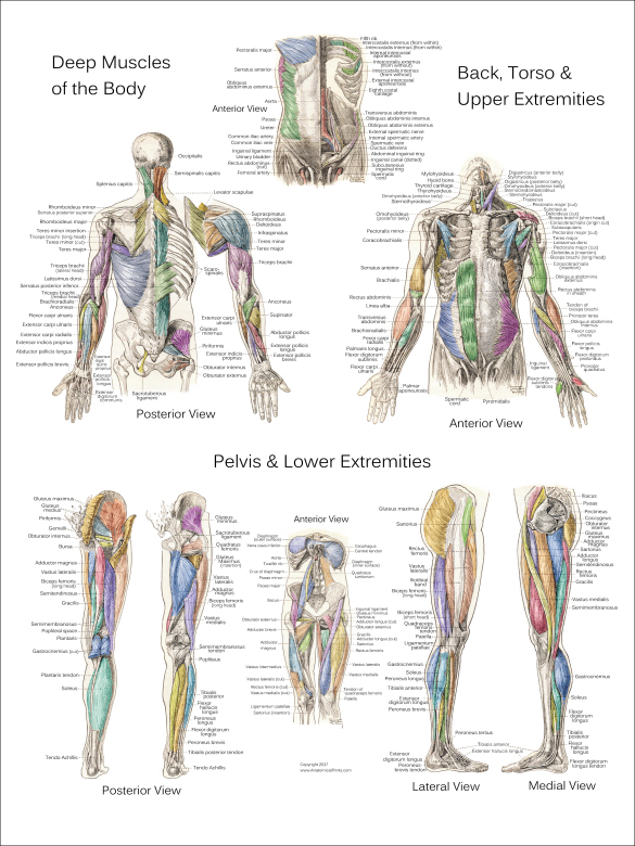
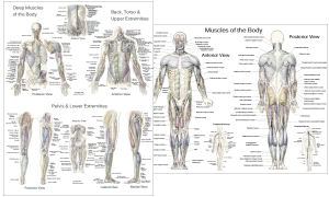
Muscle Anatomy Posters - Anterior, Posterior & Deep Layers
The Anatomy of the Lower Leg Muscles - Verywell Health The lower leg comprises two very strong, long bones: the fibula the tibia (shinbone). The tibia is stronger and more prominent than the fibula. It is located toward the middle of the lower leg. The fibula, or calf bone, is smaller and located on the lower leg's outside. The lower leg is also home to nerve fibers, including the superficial ...
Surface Muscle Anatomy Upper Extremities Arms
The Biomechanics of the Human Lower Extremity | Basic Biomechanics, 8e ... In contrast, the lower extremity is well equipped for its functions of weight bearing and locomotion. Beyond these basic functions, activities such as kicking a field goal in football, performing a long jump or a high jump, and maintaining balance en pointe in ballet reveal some of the more specialized capabilities of the lower extremity. This ...

Muscle Anatomy Posters in Spanish
Lower limb anatomy: Bones, muscles, nerves, vessels | Kenhub Therefore, try to keep them in top physical condition by giving them plenty of exercise. The lower extremity can be divided into several parts or regions, as follows: Hip Thigh Knee Leg Ankle Foot In this topic page, we will take a brief look at all of them and cover the basics of the entire lower limb. Contents Hip and pelvis Bones Muscles

Anatomy of the Skeletal System | Doctor Stock
Anatomy, Appendicular Skeleton - StatPearls - NCBI Bookshelf Clavicle Scapula Arm Humerus Forearm Radius Ulna Wrist or carpal bones Scaphoid Lunate Triquetrum Pisiform Trapezium Trapezoid Capitate Hamate Hand Metacarpals x5 Phalanx x14 Lower Limb Pelvic girdle (hip or coxal bone) Ilium Ischium Pubis Thigh Femur Leg Tibia Fibula Tarsal bones Talas Calcaneus Cuboid Medial, intermediate, and lateral cuneiform
Post a Comment for "45 lower extremities labeled"