39 microscope diagram labelled
What is skin? The layers of human skin - YouTube The skin is the largest organ of the human body, weighing approximately 16% of our bodyweight. Skin consists of multiple layers, epidermis, dermis and hypode... Labeled Microscope and Basics of Life Diagram | Quizlet PLAY. A microscope is an instrument widely to magnify and resolve the image of an object that is otherwise invisible to naked eye. For resolving the details of objects, which otherwise cannot be achieved by naked eye, a microscope is used. This set of flash cards will help the student to identify the different parts and function of the microscope.
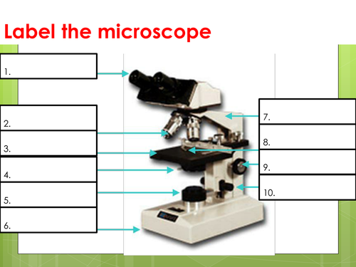
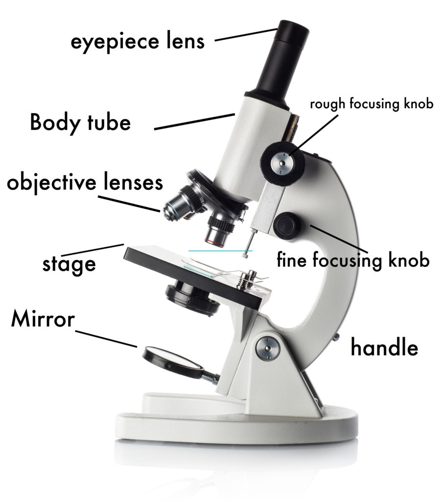
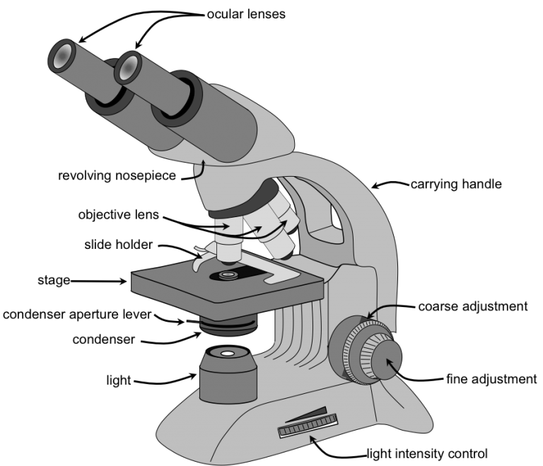
Compound Microscope Parts, Functions, and Labeled Diagram Common compound microscope parts include: Eyepiece (ocular lens) with or without Pointer: The part that is looked through at the top of the compound microscope. Eyepieces typically have a magnification between 5x & 30x. Monocular or Binocular Head: Structural support that holds & connects the eyepieces to the objective lenses.

Microscope diagram labelled
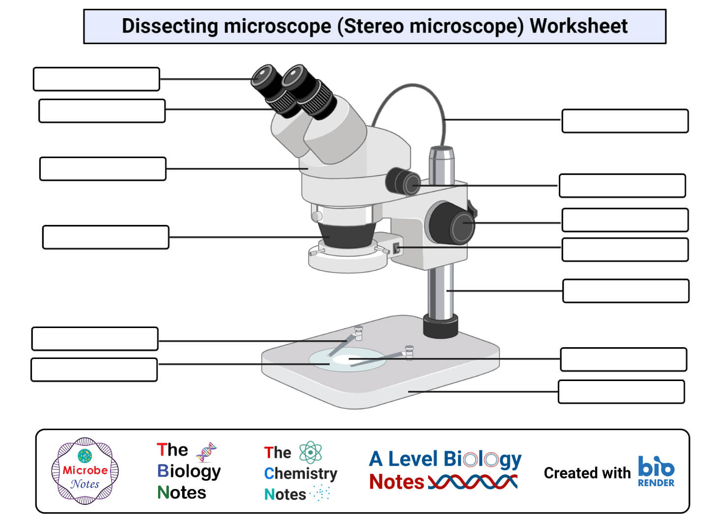
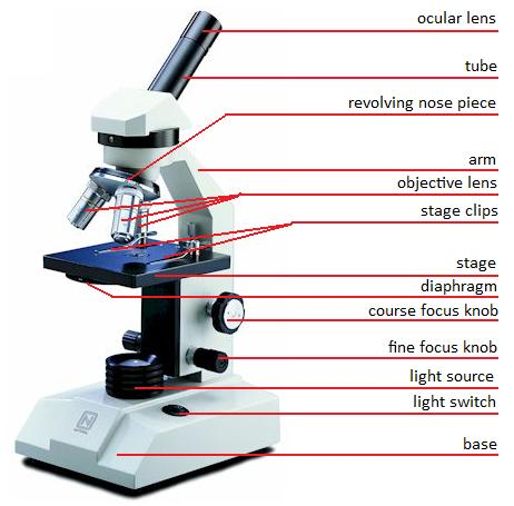
Microscope Parts, Function, & Labeled Diagram - slidingmotion Microscope parts labeled diagram gives us all the information about its parts and their position in the microscope. Microscope Parts Labeled Diagram The principle of the Microscope gives you an exact reason to use it. It works on the 3 principles. Magnification Resolving Power Numerical Aperture. Parts of Microscope Head Base Arm Eyepiece Lens Parts of Stereo Microscope (Dissecting microscope) - labeled diagram ... Labeled part diagram of a stereo microscope Major structural parts of a stereo microscope. There are three major structural parts of a stereo microscope. The viewing Head includes the upper part of the microscope, which houses the most critical optical components, including the eyepiece, objective lens, and light source of the microscope. Light microscopes - Cell structure - Edexcel - BBC Bitesize The compound microscope uses two lenses to magnify the specimen: the eyepiece and an objective lens. In most microscopes, there is a choice of objectives to use. Magnification can therefore be ...
Microscope diagram labelled. ZEISS LSM 980 with Airyscan 2 – Confocal Microscope with … Mouse brain cerebellum labelled with anti-calbinding (Alexa-568) and anti-GFAP (Alexa-488). The fluorophores were both excited with the 2-Photon laser at 780 nm and the emission spectra were simultaneously collected by the BIG.2 detector. 3D Tilling and Stitching were used to cover whole structure, and an orthogonal projection was created in ... Microscope Diagram Labeled, Unlabeled and Blank - Pinterest Microscope Diagram Labeled, Unlabeled and Blank | Parts of a Microscope - Tim's Printables. Print a microscope diagram, microscope worksheet, or practice microscope quiz in order to learn all the parts of a microscope. Tim's Printables. 38k followers . Life Science Middle School ... Label the Microscope Diagram | Download Scientific Diagram - ResearchGate Download scientific diagram | Label the Microscope Diagram from publication: Laboratory Exercises in Microbiology: Discovering the Unseen World through Hands-on Investigation | Microbiology ... Compound Microscope- Definition, Labeled Diagram, Principle, Parts, Uses The optical microscope often referred to as the light microscope, is a type of microscope that uses visible light and a system of lenses to magnify images of small subjects. There are two basic types of optical microscopes: Simple microscopes. Compound microscopes. The term "compound" in compound microscopes refers to the microscope having ...
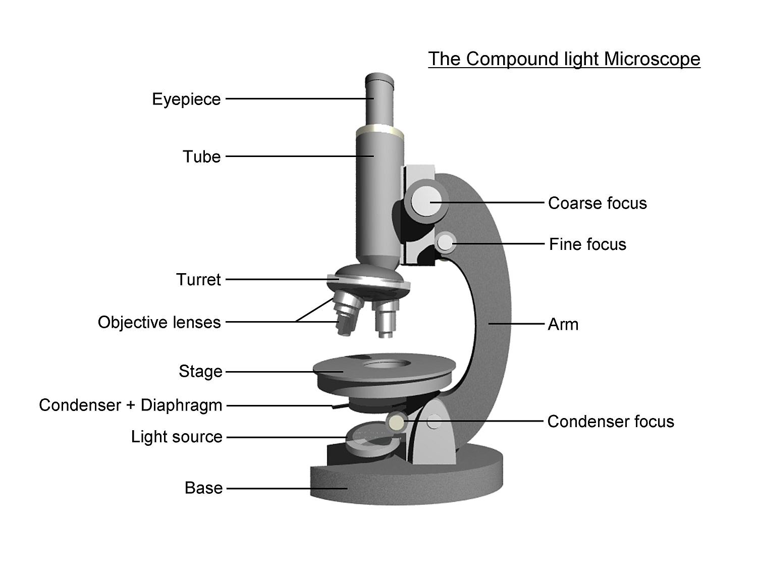
Microscope Parts and Functions Microscope Parts and Functions With Labeled Diagram and Functions How does a Compound Microscope Work?. Before exploring microscope parts and functions, you should probably understand that the compound light microscope is more complicated than just a microscope with more than one lens.. First, the purpose of a microscope is to magnify a small object or to magnify the fine details of a larger ... Label Microscope Diagram - EnchantedLearning.com Using the terms listed below, label the microscope diagram. arm - this attaches the eyepiece and body tube to the base. base - this supports the microscope. body tube - the tube that supports the eyepiece. coarse focus adjustment - a knob that makes large adjustments to the focus. diaphragm - an adjustable opening under the stage, allowing ... Parts of a microscope with functions and labeled diagram - Microbe Notes Figure: Diagram of parts of a microscope. There are three structural parts of the microscope i.e. head, base, and arm. Head - This is also known as the body. It carries the optical parts in the upper part of the microscope. Base - It acts as microscopes support. Compound Microscope: Definition, Diagram, Parts, Uses, Working ... - BYJUS Compound microscope is a type of optical microscope that is used for obtaining a high-resolution image. There are more than two lenses in a compound microscope. Learn about the working principle, parts and uses of a compound microscope along with a labeled diagram here.
Required practical - using a light microscope - BBC Bitesize Record the microscope images using labelled diagrams or produce digital images. When first examining cells or tissues with low power, draw an image at this stage, even if going on to examine the ... A Study of the Microscope and its Functions With a Labeled Diagram ... A Study of the Microscope and its Functions With a Labeled Diagram To better understand the structure and function of a microscope, we need to take a look at the labeled microscope diagrams of the compound and electron microscope. These diagrams clearly explain the functioning of the microscopes along with their respective parts. pE-300white | LED Microscope Illuminator - CoolLED CoolLED Ltd supply LED Microscope Illuminator such as pE-300white for distributors throughout the world. Call +44 (0)1264 323040(UK) or 1.800.877.0128(USA). ... Capturing high-speed multi-labelled events with LED illumination systems; ... CoolLED pE-300 white Exploded Diagram; CoolLED pE-300 Series and pE-340 fura Declaration of Conformity; Top 16 Techniques Used in Cell Biology (With Diagram) ADVERTISEMENTS: The following points highlight the top sixteen techniques used in cell biology. Some of the techniques are: 1. Immunofluorescence Microscopy 2. Ion-Exchange Chromatography 3. Affinity Chromatography 4. Partition and Adsorption Chromatography 5. Gel Filtration Chromatography 6. Radioactive Tracer Technique 7. Radioimmunoassay (RIA) 8. …
Compound Microscope Parts, Functions, and Labeled Diagram ...
Parts of the Microscope with Labeling (also Free Printouts) 5. Knobs (fine and coarse) By adjusting the knob, you can adjust the focus of the microscope. The majority of the microscope models today have the knobs mounted on the same part of the device. Image 5: The circled parts of the microscope are the fine and coarse adjustment knobs. Picture Source: bp.blogspot.com.
Microscopes: Labelling of light microscopes and difference ...
Label Microscope Diagram - EnchantedLearning.com Using the terms listed below, label the microscope diagram. arm - this attaches the eyepiece and body tube to the base. base - this supports the microscope. body tube - the tube that supports the eyepiece. coarse focus adjustment - a knob that makes large adjustments to the focus. diaphragm - an adjustable opening under the stage, allowing ...

Biology 4 U on Twitter: "Try this labelled diagram Quiz on ...
Parts of a Compound Microscope and Their Functions - NotesHippo Compound microscope mechanical parts (Microscope Diagram: 2) include base or foot, pillar, arm, inclination joint, stage, clips, diaphragm, body tube, nose piece, coarse adjustment knob and fine adjustment knob.. Base: It's the horseshoe-shaped base structure of microscope.All of the other components of the compound microscope are supported by it. ...
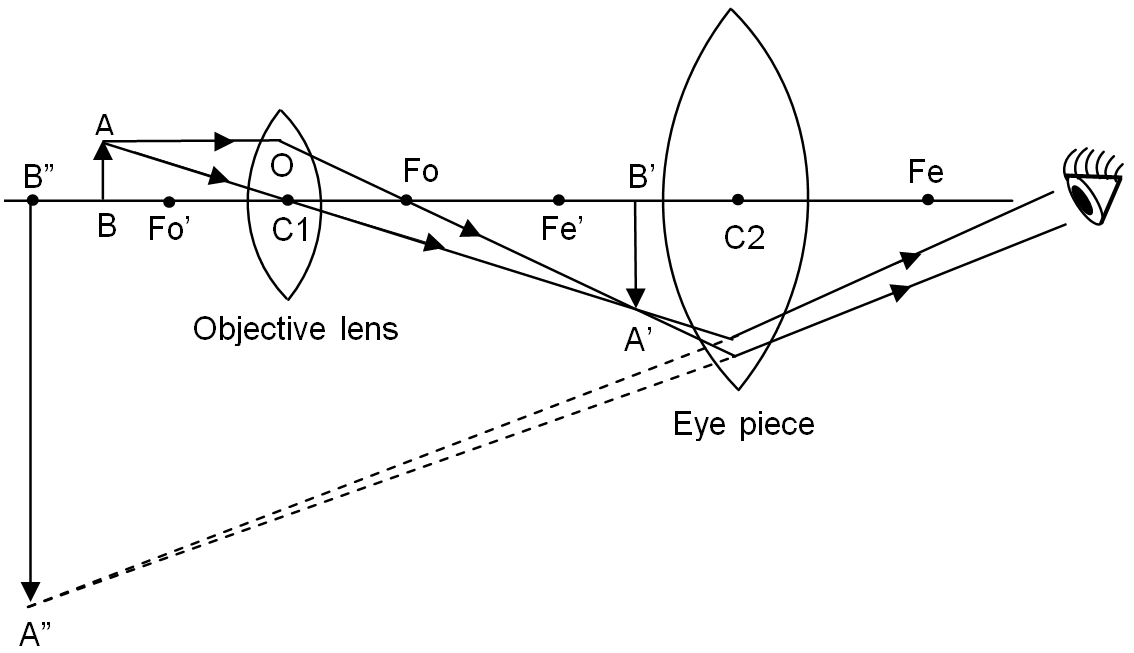
Draw a labelled ray diagram of a compound microscope and ...
Investigating cells with a light microscope - Cell structures - OCR ... Record microscope images using labelled diagrams - or you could produce digital images. When first examining cells or tissues with low power, draw an image at this stage, even if going on to ...

Microscope Labelling Review Diagram | Quizlet
Neuron under Microscope with Labeled Diagram - AnatomyLearner Neuron under microscope labelled diagram. Throughout this article, you got the different neurons labelled diagrams. Here, you will also find the diagrams of different neuron types under a microscope. The neuron diagram shows the different parts (axon, dendrites, and cell body) of the neurons.

Draw a labelled diagram of a compound microscope.
Labelled Diagram of Compound Microscope The below mentioned article provides a labelled diagram of compound microscope. Part # 1. The Stand: The stand is made up of a heavy foot which carries a curved inclinable limb or arm bearing the body tube. The foot is generally horse shoe-shaped structure (Fig. 2) which rests on table top or any other surface on which the microscope in kept.
Parts of a microscope with functions and labeled diagram
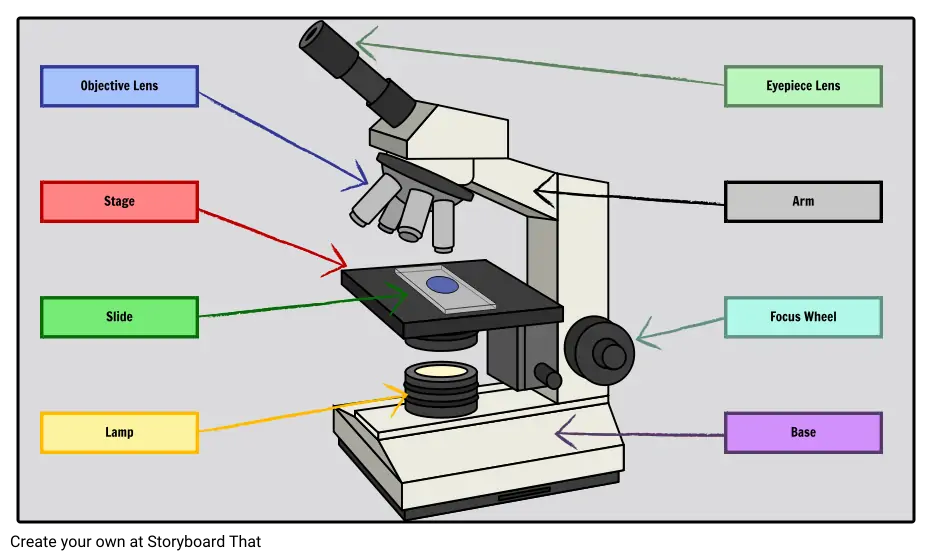
Compound Microscope Parts - Labeled Diagram and their Functions There are two major optical lens parts of a microscope: Eyepiece (10x) and Objective lenses (4x, 10x, 40x, 100x). Total magnification power is calculated by multiplying the magnification of the eyepiece and objective lens. The illuminator provides a source of light. The light is focused by the condenser and passing through the specimen placed ...

Parts of a Microscope - SmartSchool Systems
Microscope, Microscope Parts, Labeled Diagram, and Functions The description given below summarize the brief description of microscope parts used to visualize the microscopic specimens such as animal cells, plant cells, microbes, bacteria, viruses, microorganisms etc. The Microscopes parts divided into three different structural parts Head, Base, and Arms. Head/Body: It contain the optical parts in the ...

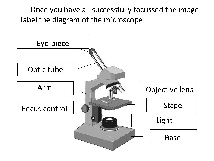
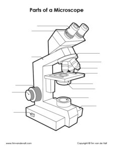
Lable the microscope worksheet
PDF Label parts of the Microscope: Answers Label parts of the Microscope: Answers Coarse Focus Fine Focus Eyepiece Arm Rack Stop Stage Clip . Created Date: 20150715115425Z ...
Labelled Diagram of Microscope Parts
Labeling the Parts of the Microscope | Microscope World Resources Labeling the Parts of the Microscope. This activity has been designed for use in homes and schools. Each microscope layout (both blank and the version with answers) are available as PDF downloads. You can view a more in-depth review of each part of the microscope here.
simple light microscope labeled - Clip Art Library
Label the microscope — Science Learning Hub All microscopes share features in common. In this interactive, you can label the different parts of a microscope. Use this with the Microscope parts activity to help students identify and label the main parts of a microscope and then describe their functions. Drag and drop the text labels onto the microscope diagram.
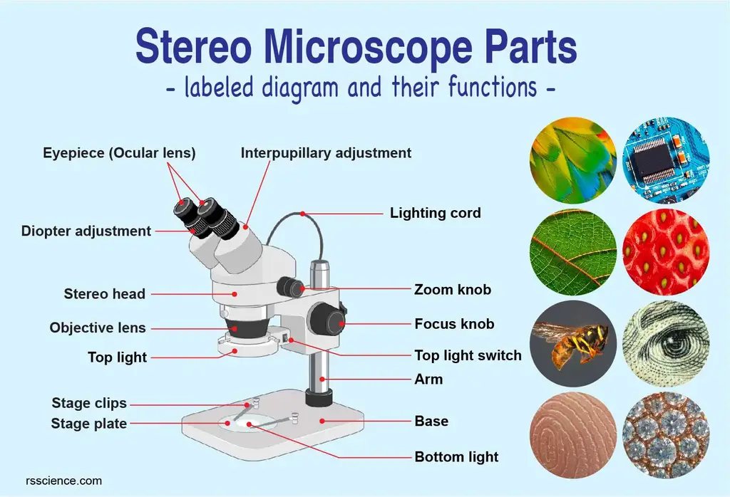
Parts of Stereo Microscope (Dissecting microscope) – labeled ...
Microscope Diagram Labeled, Unlabeled and Blank - Pinterest Dec 15, 2017 - Print a microscope diagram, microscope worksheet, or practice microscope quiz in order to learn all the parts of a microscope. Pinterest. Today. Explore. ... Microscope Diagram Labeled, Unlabeled and Blank | Parts of a Microscope - Tim's Printables. Print a microscope diagram, microscope worksheet, or practice microscope quiz ...

Diagram Carl Zeiss Mikroskop Optik, Diagram Lembar Kerja ...
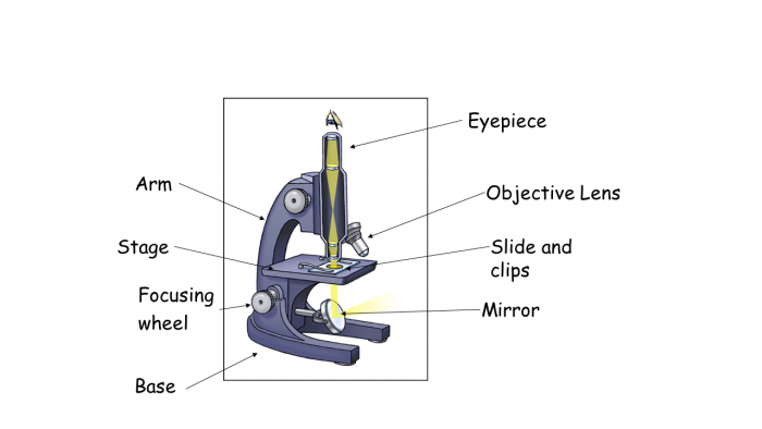
Simple Microscope - Diagram (Parts labelled), Principle, Formula and Uses Simple microscope is a magnification apparatus that uses a combination of double convex lens to form an enlarged, erect image of a specimen. The working principle of a simple microscope is that when a lens is held close to the eye, a virtual, magnified and erect image of a specimen is formed at the least possible distance from which a human eye ...
diagram picture of microscope - Clip Art Library
Microscope Labeling Diagram | Quizlet Focus and magnify light in differing amounts to view the specimen. Stage Clips. Hold the slide in place on the stage. Nosepiece. Holds the objective lenses and allows the lenses to rotate for viewing. Stage. Supports the slide where the specimen is being viewed. Lamp. Projects or reflects light upward through the diaphragm.
Microscope Using a microscope I have developed my
Compound Microscope – Diagram (Parts labelled), Principle and … Feb 03, 2022 · See: Labeled Diagram showing differences between compound and simple microscope parts Structural Components. The three structural components include. 1. Head. This is the upper part of the microscope that houses the optical parts. 2. Arm . This part connects the head with the base and provides stability to the microscope.
How to Use a Microscope
Electron microscope - Wikipedia An electron microscope is a microscope that uses a beam of accelerated electrons as a source of illumination. As the wavelength of an electron can be up to 100,000 times shorter than that of visible light photons, electron microscopes have a higher resolving power than light microscopes and can reveal the structure of smaller objects.. Electron microscopes use shaped magnetic …

Compound Microscope: Parts of Compound Microscope
Microscope Types (with labeled diagrams) and Functions Is used to view samples that are not visible to the naked eye. Uses two types of lenses - Objective and ocular lenses. Has a higher level of magnification - Typically up to 2000x. Is used in hospitals and forensic labs by scientists, biologists and researchers to study micro organisms. Compound microscope labeled diagram.

Labelling a Microscope Diagram | Quizlet
The Cell - ScienceQuiz.net The green part of the cell labelled X in the diagram is known as? a chloroplast? cytoplasm? the cell membrane? the cell nucleus; A group of similar cells that have a shared function is known as ... The diagram shows a plant cell as seen under a microscope. Two of the labels are incorrect. What are they?? Vacuole and chloroplast? Vacuole and ...
Microscope Parts Diagram PDF - Science Printables
The Parts of a Microscope (Labeled) Printable - TeacherVision The Parts of a Microscope (Labeled) Printable. Download. Add to Favorites. Share. This diagram labels and explains the function of each part of a microscope. Use this printable as a handout or transparency to help prepare students for working with laboratory equipment. Grade:

Microscope World Blog: Labeling the Parts of the Microscope
4 Major Phases of the Cell Cycle (With Diagram) - Biology … The following points highlight the four major phases of the cell cycle. The phases are: 1. G 1 (gap1) phase 2. S (synthesis) phase 3. G 2 (gap 2) phase 4. M (mitosis) phase. Cell Cycle: Phase # 1. G 1 Phase: . The G 1 phase is set in immediately after the cell division. It is characterised by a change in the chromosome from the condensed mitotic state to the more extended interphase …

Labeled Microscope Diagram | Microscope parts, Science fair ...
Microscope labeled diagram - SlideShare Microscope labeled diagram 1. The Microscope Image courtesy of: Microscopehelp.com Basic rules to using the microscope 1. You should always carry a microscope with two hands, one on the arm and the other under the base. 2. You should always start on the lowest power objective lens and should always leave the microscope on the low power lens ...
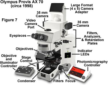
Anatomy of a Microscope | Microscopy Primer | Olympus LS
Light microscopes - Cell structure - Edexcel - BBC Bitesize The compound microscope uses two lenses to magnify the specimen: the eyepiece and an objective lens. In most microscopes, there is a choice of objectives to use. Magnification can therefore be ...
Microscope Parts Diagram - ClipArt Best
Parts of Stereo Microscope (Dissecting microscope) - labeled diagram ... Labeled part diagram of a stereo microscope Major structural parts of a stereo microscope. There are three major structural parts of a stereo microscope. The viewing Head includes the upper part of the microscope, which houses the most critical optical components, including the eyepiece, objective lens, and light source of the microscope.
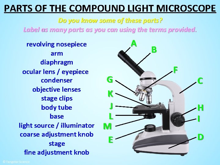
MICROSCOPE PARTS PARTS OF THE COMPOUND LIGHT MICROSCOPE
Microscope Parts, Function, & Labeled Diagram - slidingmotion Microscope parts labeled diagram gives us all the information about its parts and their position in the microscope. Microscope Parts Labeled Diagram The principle of the Microscope gives you an exact reason to use it. It works on the 3 principles. Magnification Resolving Power Numerical Aperture. Parts of Microscope Head Base Arm Eyepiece Lens
Label Microscope Diagram

Microscope Labeled Parts - ClipArt Best

This is a common compound microscope. Label its parts from A ...

Microscope labeling 2 Diagram | Quizlet
Label a microscope - Teaching resources
Label microscope - Teaching resources

Dissecting Stereo Microscope Parts and Functions

Microscope Labeling
22 Parts Of a Microscope With Their Function And Labeled ...

Simple Microscope - Diagram (Parts labelled), Principle ...

Types of Microscopes: Definition, Working Principle, Diagram ...

sam on Twitter: "WhatsApp: All the science emoji ranking ...

Diagram of traveling microscope setup with implant cast and ...
Revise the Parts of a Microscope Worksheet - EdPlace
Labeled Microscope Storyboard by oliversmith
Post a Comment for "39 microscope diagram labelled"