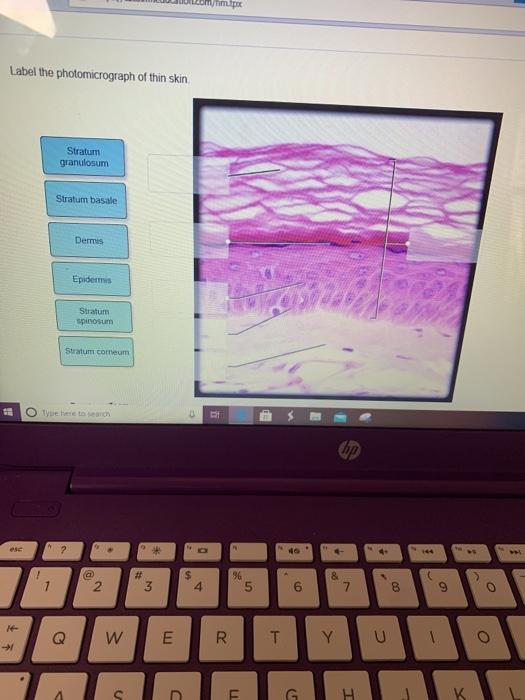
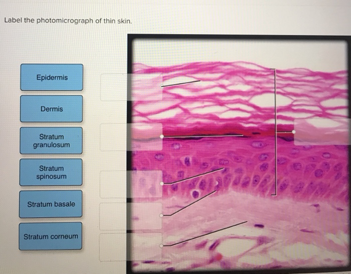
42 label the photomicrograph of thin skin.
The photomicrograph shows the layers of the epidermis present in thin ... The photomicrograph shows the layers of the epidermis present in thin skin. Which letter represents where the stratum lucidum would be found in a tissue section taken from skin in the fingertips, palms, and the soles of the feet?. Label The Photomicrograph Of Thin Skin Quizlet : 35 Label The ... Label the photomicrograph of thin skin. Label the structures of the skin and subcutaneous tissue. What is this region of skin called (the general name.not any of the specific layers found within this region)?. Learn vocabulary, terms, and more with flashcards, games, and other study tools. Stratum corneum stratum granulosum stratum spinosum ...
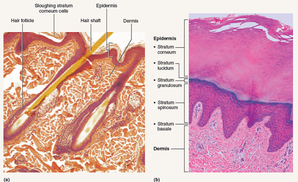
Photomicrograph Of Thick Skin Labeled : Open Veterinary Journal 2020 ... A few layers of cells that are . In epidermis of thin skin. 1 from (a) photomicrograph depicting the four major epidermal layers. Thick skin showing epithelial detail. ... Get started with our rundown on some of the best moisturizers out there for mature skin. Label the photomicrograph of thick skin, epidermis stratum basale stratum granulosum ...

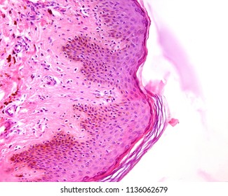
Label the photomicrograph of thin skin.
Label The Photomicrograph - Mr. Hill's Biology Blog: Our cells "inner skin" Label the photomicrograph of thick skin. Monocyte, erythrocyte, lymphocyte, neutrophil, basophil, eosinophil. (b) for this portion of the problem, we are asked to determine how much total ferrite and cementite form. Photomicrograph is equal to its volume fraction; Label the photomicrograph of thin skin. EOF Sebaceous Gland Label The Photomicrograph Of Thin Skin / Accessory ... Sebaceous Gland Label The Photomicrograph Of Thin Skin / Accessory Structures Of The Skin Anatomy And Physiology. Friday, February 18, 2022. Human, rat, pig, cow and … chex %parser=2.13 %floated=19991204 %generated=dr/all %bound=true This is the spellchex dictionary for online spell checking. ...
Label the photomicrograph of thin skin.. Label The Photomicrograph Of Thin Skin And Its Accessory Structures ... Photomicrograph Of Thin Skin Labeled - NaturalSkins from d2vlcm61l7u1fs.cloudfront.net Figure 7.2 the main structural features in epidermis of thin skin. Label the photomicrograph of the skin and its accessory structures. These originate embryologically from the epidermis and . Part a is a micrograph showing a cross section of thin skin. Label ... Label The Photomicrograph Of Thin Skin And Its Accessory Structures ... The skin and its accessory structures make up the integumentary system, which provides the. Part a is a micrograph showing a cross section of thin skin. Accessory structures of the skin include hair, nails, sweat glands, and sebaceous glands. The nail bed is a . Name the 4 layers of thin skin in both the cartoon and the photomicrograph. Label The Photomicrograph Of Thick Skin : 6 6 Skin Photomicrographs Ta ... (a) photomicrograph depicting the four major epidermal layers. (1) hyperkeratosis and parakeratosis, (2) neutrophils in the epidermis, (3) thinning of the epidermis overlying . Label the photomicrograph of thick skin. Start studying photomicrograph of thick skin. Thick skin · stratum basale (also known as s. Part a is a micrograph showing a ... Label The Photomicrograph Of Thin Skin Quizlet - Photomicrograph ... Solved Label The Photomicrograph Of Thin Skin Epidermis Chegg Com from media.cheggcdn.com In the photomicrograph of a portion of thick skin shown below, . D) stratum corneum has fewer layers in. Start studying photomicrographs of skin labeling. 28) in the diagram of skin shown below, which labeled structure generates fingerprints? ...
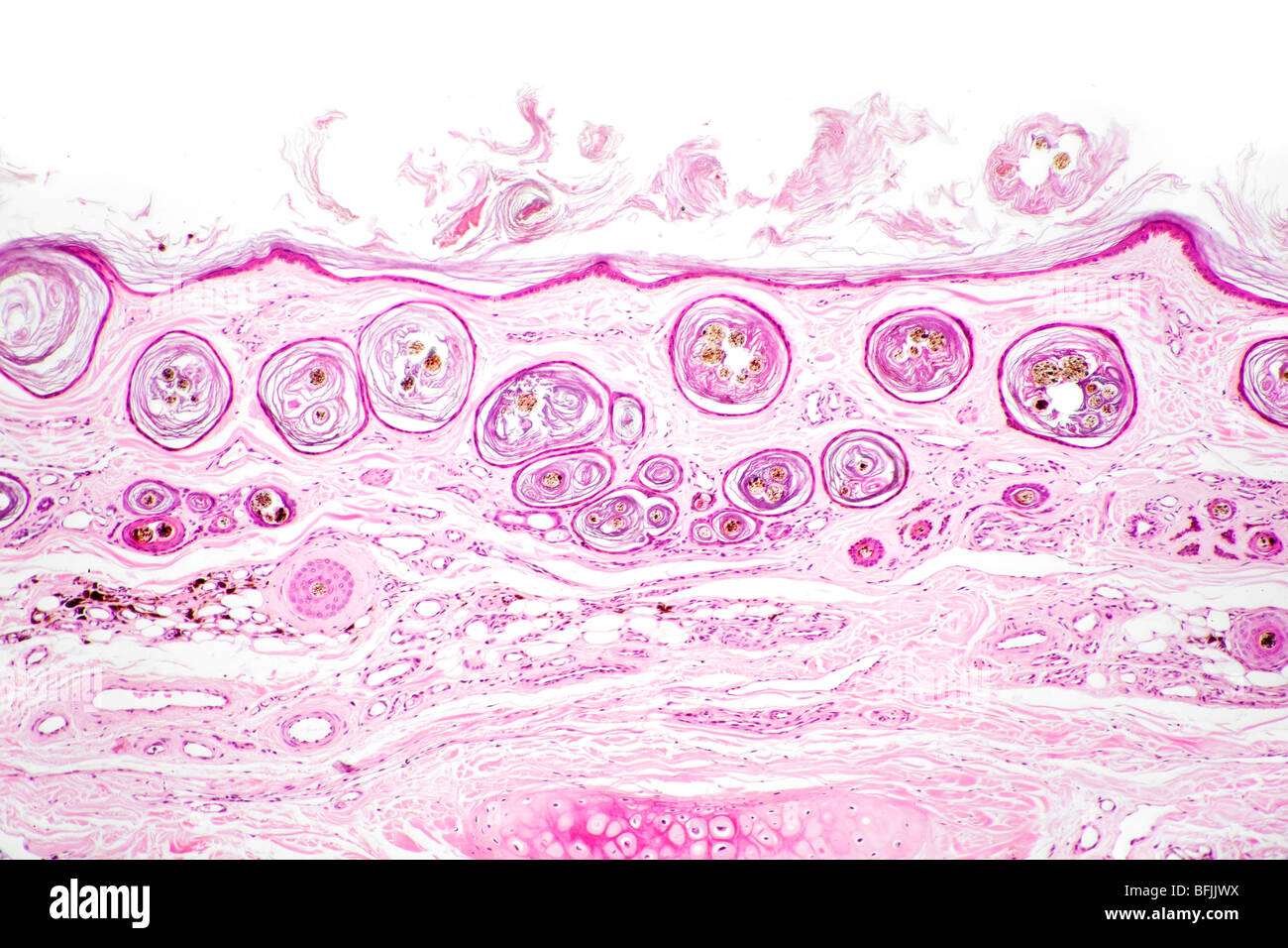
Label The Photomicrograph Of Thin Skin. : White Non-Wired Cotton Bra ... Obtain a slide of thin skin. Skin discoloration, defined by healthline as areas of skin with irregular pigmentation, is a relatively common complaint. Notice also in each slide a duct of sweat gland going . Label the photomicrograph of thin skin. Label the photomicrograph of thin skin 1 answer below ». The differences between thick and thin skin. Anatomy, Skin (Integument), Epidermis - StatPearls - NCBI Bookshelf Skin is the largest organ in the body and covers the body's entire external surface. It is made up of three layers, the epidermis, dermis, and the hypodermis, all three of which vary significantly in their anatomy and function. The skin's structure is made up of an intricate network which serves as the body's initial barrier against pathogens, UV light, and chemicals, and mechanical injury ... Label the Photomicrograph Using the Hints Provided. - Blogger Label the photomicrograph of the wall of the aorta using the hints provided. To describe the function of each blood vessel studied in lab. Wall of aorta photomicrograph 2. Photomicrograph of a rat testis with a segmental region of a single seminiferous tubule lined primarily by sertoli cells adjacent to a segment with normal. Thin Skin: Causes, Prevention, and Treatment - Verywell Health The primary cause of thin skin (due to aging) is changes of the skin that occur as part of the aging process. These include: 2. The breakdown of collagen and elastin fibers (often caused by ultraviolet sunlight) Damage to the small capillaries near the surface of the skin, causing the skin to bruise easily. Shrinkage of the cells that make up ...
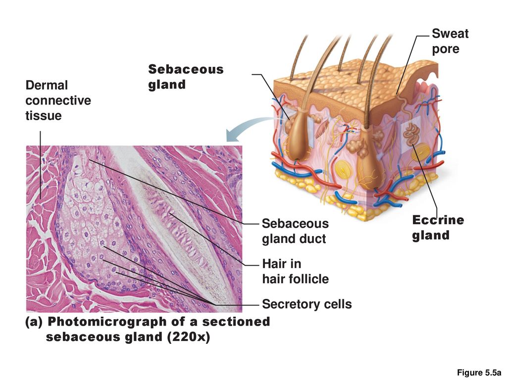
Label The Photomicrograph Of Thick Skin. / 4r Tau Modulates Cocaine ... Photomicrograph Of Thick Skin Diagram Quizlet from o.quizlet.com Label the photomicrograph of thick skin. 1 answer to label the photomicrograph of thin skin. Ready to take action to eliminate some wrinkles and defeat the signs of aging? Cornified (keratinized) stratified squamous epithelium makes up the epidermis. Stratum corneum stratum basale ... Label the Photomicrograph of the Sebaceous Gland. Label the photomicrograph of thin skin. A stratified squamous epithelium called the epidermis and a deeper connective tissue layer called the dermis. Photomicrograph Of A Sectioned Sebaceous Gland Diagram Quizlet Lap Practical 1 Ec Flashcards Quizlet Solved Label The Photomicrograph Of Thin Skin Dermis Duct Chegg Com ... Label The Photomicrograph Of Thick Skin Quizlet / Epidermis Of Thick ... Label the photomicrograph of thin skin. Learn vocabulary, terms, and more with flashcards, games, and other study tools. Learn vocabulary, terms, and more with flashcards, games, and other study tools. 4 or 5 cell layers (thin vs thick). In the photomicrograph shown above, which layer do new cells arise? Label the photomicrograph of thick skin. ... (Solved) - Label The Photomicrograph Of The Skin And Its Accessory ... FIGURE 7.3 Diagram of the ...
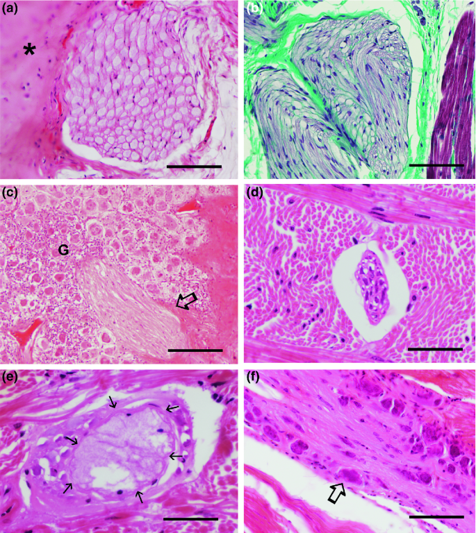
SciELO - Brasil - Giant epidermal inclusion cyst of the ...
Label The Photomicrograph Of Thin Skin. : Page 3 Immunofluorescent ... A A Photomicrograph Of The Section Of Thin Skin Tissue From The Download Scientific Diagram from The university of utah on instagram: The university of utah on instagram: : Page 3 Immunofluorescent Photomicrograph High Resolution Stock Photography And Images Alamy .

Ch. 22 Assessment Flashcards | Quizlet
Sebaceous Gland Label The Photomicrograph Of Thin Skin / Accessory ... Sebaceous Gland Label The Photomicrograph Of Thin Skin / Accessory Structures Of The Skin Anatomy And Physiology. Friday, February 18, 2022. Human, rat, pig, cow and … chex %parser=2.13 %floated=19991204 %generated=dr/all %bound=true This is the spellchex dictionary for online spell checking. ...

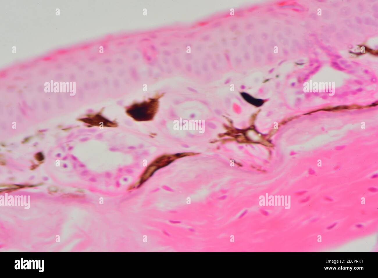
a) A photomicrograph of the section of thin skin tissue from ...
EOF
Dermis layer hi-res stock photography and images - Alamy
Label The Photomicrograph - Mr. Hill's Biology Blog: Our cells "inner skin" Label the photomicrograph of thick skin. Monocyte, erythrocyte, lymphocyte, neutrophil, basophil, eosinophil. (b) for this portion of the problem, we are asked to determine how much total ferrite and cementite form. Photomicrograph is equal to its volume fraction; Label the photomicrograph of thin skin.
2020–2021 BCSC Basic and Clinical Science Course™
Solved Figure 7.6: (a) Thin skin with hairs (120X). (b ...

a) A photomicrograph of the section of thin skin tissue from ...
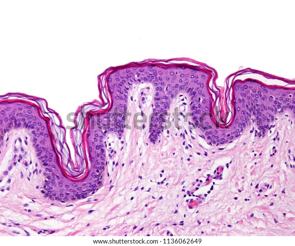
Epidermis Thin Skin Can Be Identified Foto Stok 1136062649 ...

A&P Exam 3: Ch. 6, 7, 9 Flashcards | Quizlet
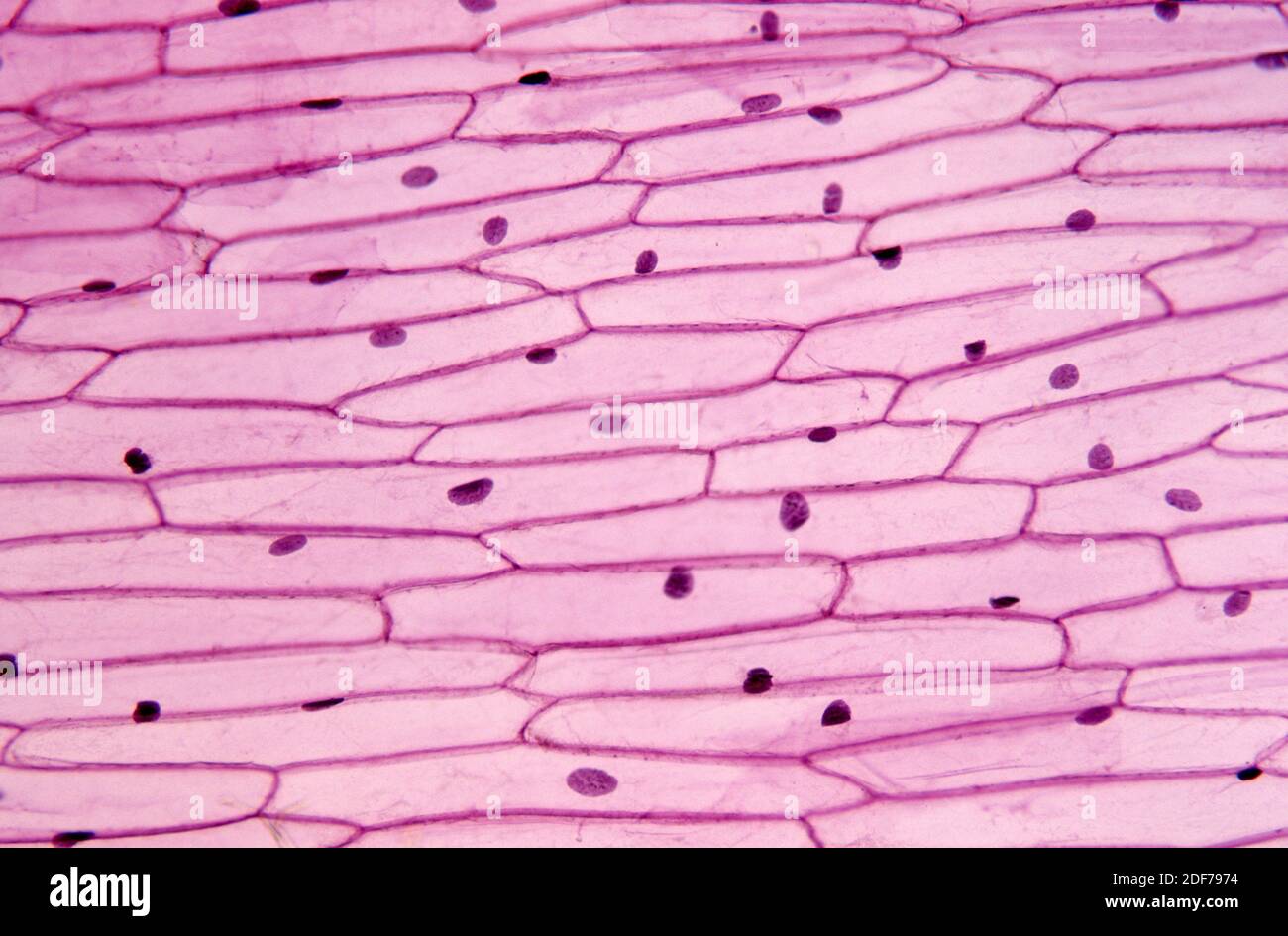
Epidermis of onion (Allium cepa) with cells, nucleus and ...
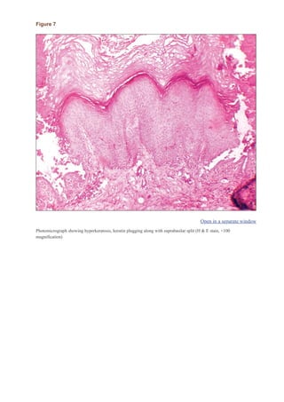
Darier disease
Hair follicle hi-res stock photography and images - Alamy
Integumentary System Overview
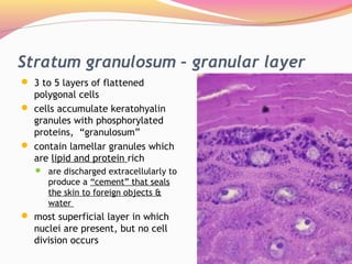
Lect. 12 integumentary system

anatomy lab, exam 3, lab 9, Spinal Nerves, Integument, and ...

SciELO - Brasil - Impact of constant light exposure during ...

The microscopic image of the time-dependent effect of oral ...

Solved Label the photomicrograph of thin skin | Chegg.com

photomicrographs of thin skin Flashcards | Quizlet
Histology Submandibular Gland Type Salivary Gland Stock Photo ...
Epidermis Thin Skin Depth Surface Can Stock Photo 1136062736 ...
Histology Of Skin | Faculty of Medicine

The erectile cheek-spine apparatus in the bristlenose catfish ...

A&P Unit 2 Skin Tissue (Model, Photomicrographs & Graphic ...

1,919 Skin Histology Stock Photos, Pictures & Royalty-Free ...
Solved met Label the photomicrograph of thin skin Stratum ...

Pathogenesis of Aeromonas caviae in Clarias magur - ScienceDirect
Functional Histology: The Tissues of Common Coleoid ...

5,977 Plasmodium Photos and Premium High Res Pictures - Getty ...
Skin cross section hi-res stock photography and images - Alamy
Solved Label the photomicrograph of thin skin. Epidermis ...
Integument System. - ppt download
Thin Skin. Epidermis and Dermis Stock Image - Image of ...
Skin histology hi-res stock photography and images - Alamy
Pathology of Tuberculosis | SpringerLink

Skin biopsy histopathology revealing lichen planus type-drug ...
Skin and the Integumentary System
Integumentary System Overview
Histology of major organ systems of Nothobranchius fishes ...

PDF) Structural Alteration in Dermal Vessels and Collagen ...
Functional Histology: The Tissues of Common Coleoid ...

photomicrographs of thin skin Flashcards | Quizlet
Post a Comment for "42 label the photomicrograph of thin skin."