44 microscope diagram to label
Interface Embrittlement Between 63Sn-37Pb Solder and Au Layer—Part 1 ... The (i) diagram shows a section of the post-solidification microstructure in (a). The Au x Sn y IMC layer formed between the Au layer and molten Sn-Pb solder. Footnote 1 Solid-state diffusion and reaction processes resulted in the development of two individual Au m Sn n and Au s Sn t IMC layers as illustrated in the (ii) diagram. Light Microscope (Theory) - Amrita Vishwa Vidyapeetham Microscope is an optical instrument that uses lens or combination of lens to produce magnified images that are too small to seen by unaided eye. Microscope provides the enlarged view that helps in examining and analyzing the image.
APA Citation Guide (7th edition) : Sample Paper, Reference List ... An annotated bibliography is a list of citations for various books, articles, and other sources on a topic. The annotated bibliography looks like a Reference page but includes an annotation after each source cited. An annotation is a short summary and/or critical evaluation of a source.
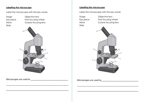
Microscope diagram to label
plant cell organelles Plant cells This basic structure of a plant cell is shown below the same plant cell as viewed with the light microscope and with the transmission electron microscope. ... Animal Cell Model Diagram Project Parts Structure Labeled Coloring And Plant Cell Organelles Cake Animal Ce Plant Cell Diagram Cell Diagram Plant Cell Labeled: Gram Stain Technique - Amrita Vishwa Vidyapeetham Wipe the glass slide with spirit and wave the slide over the Bunsen burner to remove any unwanted microorganisms in the slide. Label one side of the glass slide with 1. Your initials 2. The date While flaming the inoculation loop be sure that each segment of metal glows orange/red-hot before you move the next segment into the flame. Blood Smear - Understand the Test - Testing.com Purpose of the test. A blood smear is used to evaluate your red blood cells (RBCs), noting any abnormal differences in size, shape, or other physical appearances such as that seen in various anemias, sickle cell disease, Thalassemia, or other disorders. Evaluation of white blood cells (WBCs) is required especially if they are increased or ...
Microscope diagram to label. N-STORM | Super-Resolution Microscopes | Nikon Microscope Products ... N-STORM takes advantage of Nikon's powerful Ti2-E inverted microscope and applies high-accuracy, multi-color localization and reconstruction in three dimensions (xyz) to enable super-resolution imaging at tenfold the resolution of conventional light microscopes (up to approximately 20 nm in xy). This powerful technology enables the ... Dog Hock Anatomy with Diagram - Canine Tarsal Joint You may also find these arrangements (3 rows of dog's tarsal) in the below-mentioned diagram - Both the Table and diagram might help you to understand the number of tarsal bones with their proper arrangement. Here, three rows represent - First row (proximal) - talus or tibial tarsal (medially), and calcaneus or fibular tarsal (laterally), Prokaryote Vs Eukaryote Venn Diagram Notes - Otosection Prokaryotes vs. eukaryotes venn diagram drag drop, labeling activity in slides. this slides activity challenges students to compare and contrast prokaryotes and eukaryotes. the resource actually contains two assignments all in one.slide #1 is a drag and drop activity with 17 options, while slide #2 contains clickable text boxes which students. Plant Animal Cells Graphic Organiser - Otosection Label me! printouts. use the chart below to compare and contrast characteristics of plant and animal cells, noting whether the cell structure belongs to plant cells, animal cells, or both plant and animal cells. this is a thumbnail of the plant and animal cells graphic organizer print out. the full size printout is available only to site members.
Optical detection of multiple bacterial species using nanometer-scaled ... As a result, E. coli O26, E. coli O157, and S. aureus were observed as white, red, and blue scattered light, respectively, under the microscope. Furthermore, when adding predetermined amounts of E ... Metaphase - Genome.gov Metaphase is a stage during the process of cell division (mitosis or meiosis). Normally, individual chromosomes are spread out in the cell nucleus. During metaphase, the nucleus dissolves and the cell's chromosomes condense and move together, aligning in the center of the dividing cell. At this stage, the chromosomes are distinguishable when ... Motoneurons innervation determines the distinct gene expressions in ... Note that several nuclei are enriched at R-BTX-labeled synaptic sites (indicated with dotted lines). R-BTX (red). B Quantification of number of nucleus in the synaptic region (SR) or the non-synaptic region (NSR) in A. n = 38 myofibers. Results were from three mice. C Schematic diagram of the separation of SR and NSR. Mr. Jones's Science Class Earth, Moon, & Sun System (PPT.) Seasons Interactive (Online Activity) Moon Phases - Introductory Activity. Modeling the Phases of the Moon. Problems in Space (Online Activity) Lunar & Solar Eclipses - Webquest.
DNA - Wikipedia Deoxyribonucleic acid (/ d iː ˈ ɒ k s ɪ ˌ r aɪ b oʊ nj uː ˌ k l iː ɪ k,-ˌ k l eɪ-/ (); DNA) is a polymer composed of two polynucleotide chains that coil around each other to form a double helix carrying genetic instructions for the development, functioning, growth and reproduction of all known organisms and many viruses.DNA and ribonucleic acid (RNA) are nucleic acids. Brainpop Microscope Worksheet - Worksheet Genius Label The Parts Of The Compound Microscope. Brainpop is a subscription website that has excellent science videos, as well as history, english, math, arts, etc. Unit one the nervous system university of washington. ... The animal cell diagram is widely asked in class 10 and 12 examinations and is beneficial to understand the structure and ... A Novel Method for COVID-19 Detection Based on DCNNs and Hierarchical ... The worldwide outbreak of the new coronavirus disease (COVID-19) has been declared a pandemic by the World Health Organization (WHO). It has a devastating impact on daily life, public health, and global economy. Due to the highly infectiousness, it is urgent to early screening of suspected cases quickly and accurately. Chest X-ray medical image, as a diagnostic basis for COVID-19, arouses ... Fluorescence In Situ Hybridization (FISH) - Genome.gov The fluorescently labeled DNA finds its matching segment on one of the chromosomes, where it sticks. By looking at the chromosomes under a microscope, a researcher can find the region where the DNA is bound because of the fluorescent dye attached to it. This information thus reveals the location of that piece of DNA in the starting genome.
The Compound Light Microscope Label the following parts on ...
A comprehensive mouse brain acetylome-the cellular-specific ... Nε-lysine acetylation is a reversible posttranslational modification (PTM) involved in multiple physiological functions. Genetic and animal studies have documented the critical roles of protein acetylation in brain development, functions, and various neurological disorders. However, the underlying cellular and molecular mechanism are still partially understood. Here, we profiled and ...

Simple Microscope- Definition, Principle, Magnification ...
Dicot Stem Cross Section - TobykruwFlores Draw a labelled diagram of cross-section of a leaf. Dicot vs Monocot Stem. German Centre for Integrative Biodiversity Research iDiv Halle. In a cross-section of a dicot stem you will find an epidermis hypodermis endodermis ground tissues and vascular bundles. Multicellular epidermal hairs all over the epidermis.
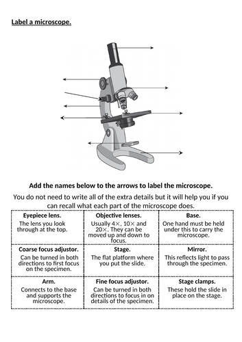
New AQA biology microscope lesson | Teaching Resources
Dicot Root Cross Section - KamorakruwWalton Pine Life History Composite - Prepared Microscope Slide - 75 x 25mm - Biology Microscopy - Eisco Labs. Half of the total solution should be sprayed in one direction and the other half sprayed perpendicular to the first application. Monocot Dicot Root Cross Section - Prepared Microscope Slide - 75 x 25mm - Biology Microscopy - Eisco Labs.

Lable the microscope worksheet
Pulmonary Artery: Anatomy, Function, and Significance - Verywell Health The main pulmonary artery is a blood vessel that comes out of the heart. It is also called the pulmonary trunk. The pulmonary artery is split into the left and right pulmonary arteries. These arteries carry blood with low oxygen and high carbon dioxide to the lungs. In the lungs, the blood gets replenished by inhaled oxygen and excess carbon ...
Lab - Microscope: MAH-Summer 2019-Anatomy and Physiology I
Antibody stabilization for thermally accelerated deep immunostaining ... a, Schematic diagram of antibody (Ab) diffusion to reach the deep tissue antigen (Ag) target.K a (T) is the association constant of the Ab-Ag binding reactions at a given temperature T, and D ...
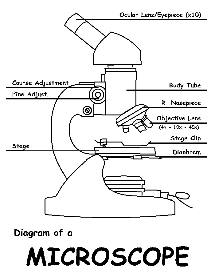
diagram - Clip Art Library
Drosophila melanogaster - Wikipedia Drosophila melanogaster is a species of fly (the taxonomic order Diptera) in the family Drosophilidae.The species is often referred to as the fruit fly or lesser fruit fly, or less commonly the "vinegar fly" or "pomace fly". Starting with Charles W. Woodworth's 1901 proposal of the use of this species as a model organism, D. melanogaster continues to be widely used for biological research in ...
7Ac Microscope Labelling Worksheet | Teaching Resources
connective tissue | Definition, Components, & Function connective tissue, group of tissues in the body that maintain the form of the body and its organs and provide cohesion and internal support. The connective tissues include several types of fibrous tissue that vary only in their density and cellularity, as well as the more specialized and recognizable variants—bone, ligaments, tendons, cartilage, and adipose (fat) tissue. In the abdominal ...
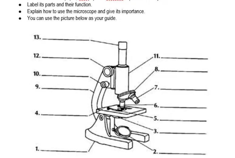
Answered: Label its parts and their function.… | bartleby
9.3: SEM and its Applications for Polymer Science The formula for magnification is shown in 9.3.1, where M is magnification, f is focal length, u is the distance between object and lens, and v is distance from lens to the image. (9.3.1) M = f u − f = v − f f Figure 9.3. 1 Basic microscope diagram illustrating inverted image and distances u, f, and v.

Parts of the Microscope with Labeling (also Free Printouts ...
What Is The Purpose Of A Microscope Microscope Parts and Functions With Labeled Diagram and Functions How does a Compound Microscope Work?. Before exploring microscope parts and functions, you should probably understand that the compound light microscope is more complicated than just a microscope with more than one lens.. First, the purpose of a microscope is to magnify a small ...

Parts of the Microscope worksheet
ECLIPSE Ti2 Series | Inverted Microscopes | Nikon Microscope Products ... The ECLIPSE Ti2 inverted microscope delivers an unparalleled 25mm field of view (FOV) that revolutionizes the way you see. With this incredible FOV, the Ti2 maximizes the sensor area of large-format CMOS cameras without making compromises, and significantly improves data throughput.

Label microscope pt.1 Diagram | Quizlet
Gear Skiving—A Step Changing Manufacturing Process Applicable to ... Figure 6 Okuma MULTUS U3000, kinematic diagram. Inspection was undertaken using a Hexagon Leitz PMM-C, which is a precision CMM with gear inspection capability. Its accuracy specification, MPE-e, was 0.023622 + L / 800 thou, where L is the measurement length, the machine has three degrees of freedom (X, Y, Z), and it was maintained to a strict ...
:max_bytes(150000):strip_icc()/microscopecolor3-58b978735f9b58af5c495abe.png)
Learn About Microscopes With Fun, Free Printables
Blood Smear - Understand the Test - Testing.com Purpose of the test. A blood smear is used to evaluate your red blood cells (RBCs), noting any abnormal differences in size, shape, or other physical appearances such as that seen in various anemias, sickle cell disease, Thalassemia, or other disorders. Evaluation of white blood cells (WBCs) is required especially if they are increased or ...

PSB Microscopy Worksheet - Microscopy Worksheet In order to ...
Gram Stain Technique - Amrita Vishwa Vidyapeetham Wipe the glass slide with spirit and wave the slide over the Bunsen burner to remove any unwanted microorganisms in the slide. Label one side of the glass slide with 1. Your initials 2. The date While flaming the inoculation loop be sure that each segment of metal glows orange/red-hot before you move the next segment into the flame.

Microscope Labeling
plant cell organelles Plant cells This basic structure of a plant cell is shown below the same plant cell as viewed with the light microscope and with the transmission electron microscope. ... Animal Cell Model Diagram Project Parts Structure Labeled Coloring And Plant Cell Organelles Cake Animal Ce Plant Cell Diagram Cell Diagram Plant Cell Labeled:

Carl Zeiss Microscopy Optical microscope Worksheet Diagram ...

Simple Microscope - Diagram (Parts labelled), Principle ...
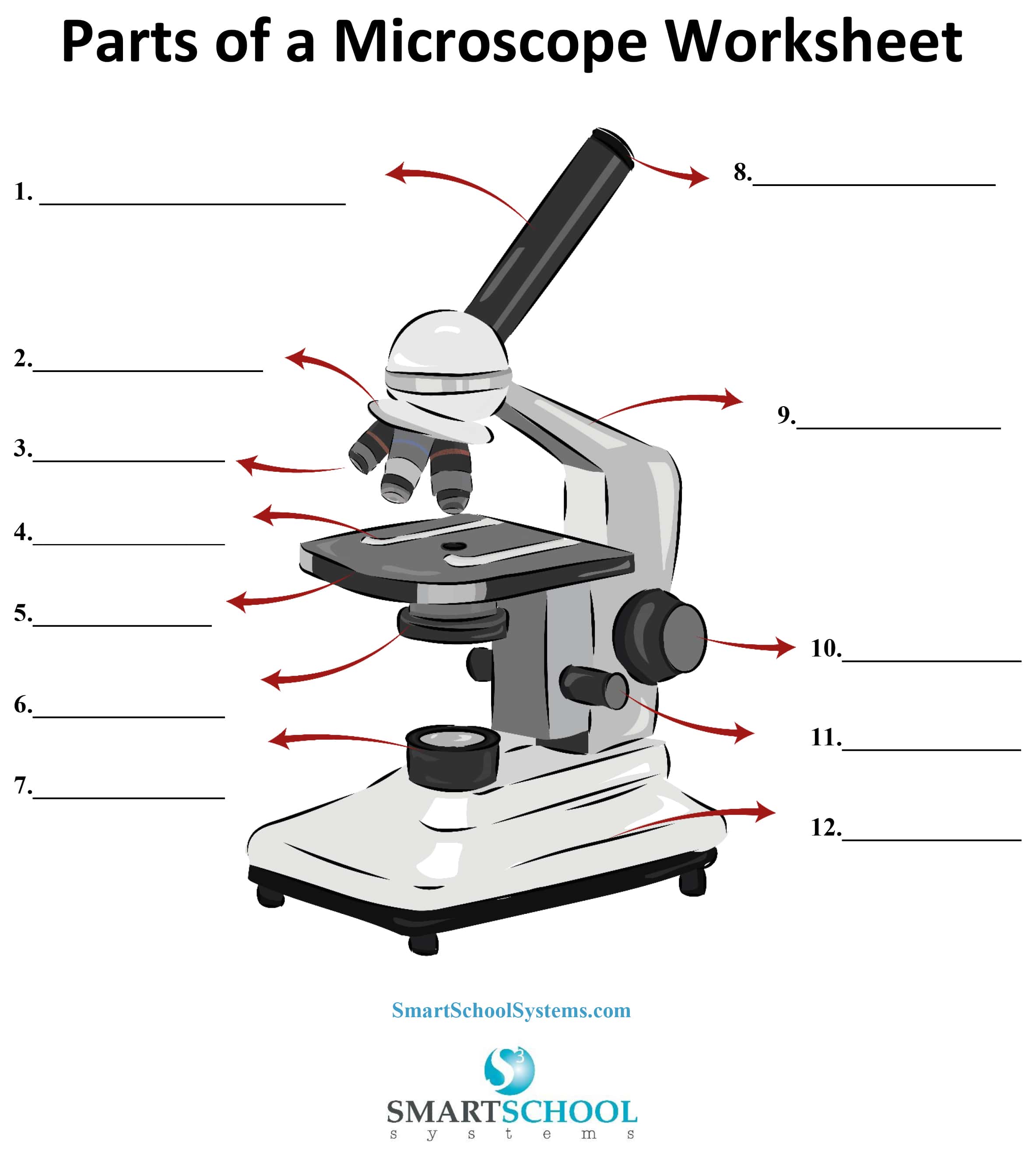
Parts of a Microscope - SmartSchool Systems

Biology - labeling a compound microscope Diagram | Quizlet
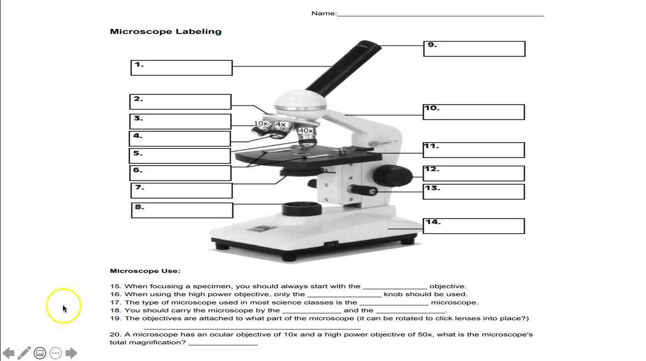
Microscope Labeling
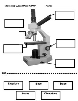
Microscope Labeling Activity

microscope drawing with label - Clip Art Library

Label the Microscope Diagram | Download Scientific Diagram
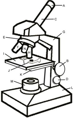
Microscope Diagram To Label - ClipArt Best

Label a Microscope Worksheet
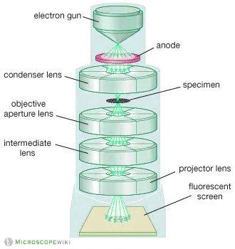
Electron Microscope Principle, Uses, Types and Images ...

MICROSCOPE Labeling - Part - 3
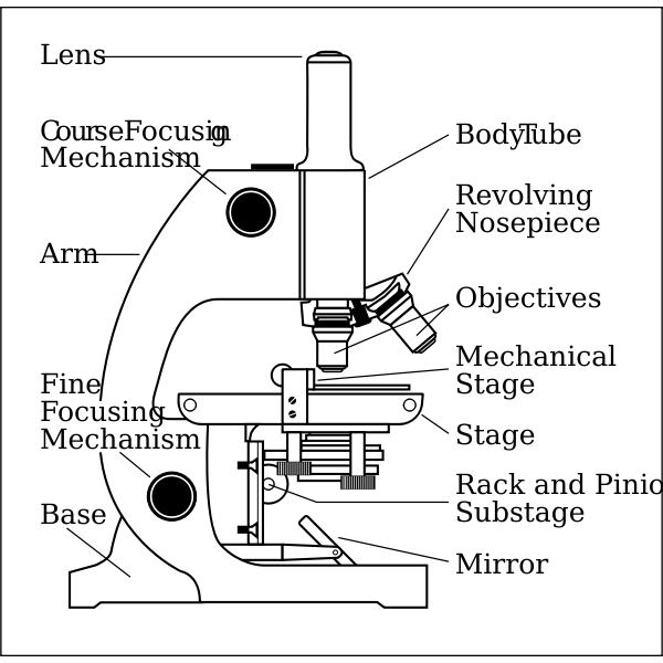
Parts of a Microscope

Dissecting Stereo Microscope Parts and Functions

Labeling a Microscope Free Worksheet Pack
Compound Microscope Parts, Functions, and Labeled Diagram ...

Compound Light Microscope Labeling Diagram | Quizlet
Name Date Sci STANDARD MICROSCOPE DIAGRAM Label only the ...
Microscope Biology - 2022
Microscope Maintenance Tips

Compound Microscope Parts – Labeled Diagram and their ...

Parts of a Microscope Microscope Basics. Label the Compound ...

Microscope Diagram Labeled, Unlabeled and Blank | Parts of a ...

Microscope worksheet

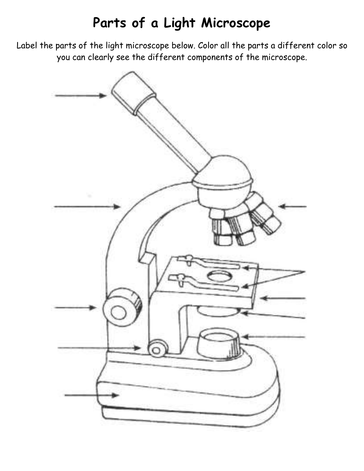
Parts of a Light Microscope Activity | Labeling Task

Compound Microscope Parts, Functions, and Labeled Diagram ...

Label the Microscope Diagram | Quizlet

Label the light microscope | Teaching Resources

Types of Microscopes: Definition, Working Principle, Diagram ...

Compound Microscope: Parts of Compound Microscope
Post a Comment for "44 microscope diagram to label"